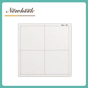
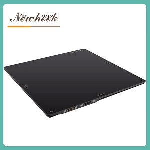
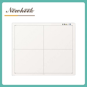
For patients with a larger body habitus, the hardest constraints usually sit on the detector side of the room. Three questions decide whether the exam is one clean exposure or a series of retakes: how much anatomy the detector’s active area actually covers, how the panel gets positioned under (or beside) the patient, and whether the panel’s handling — weight, thickness, edges, cable or battery — supports a workflow where every extra move costs more. The tube, table and generator matter too, but this article stays on the detector side: coverage, positioning, and the questions worth asking before you buy.
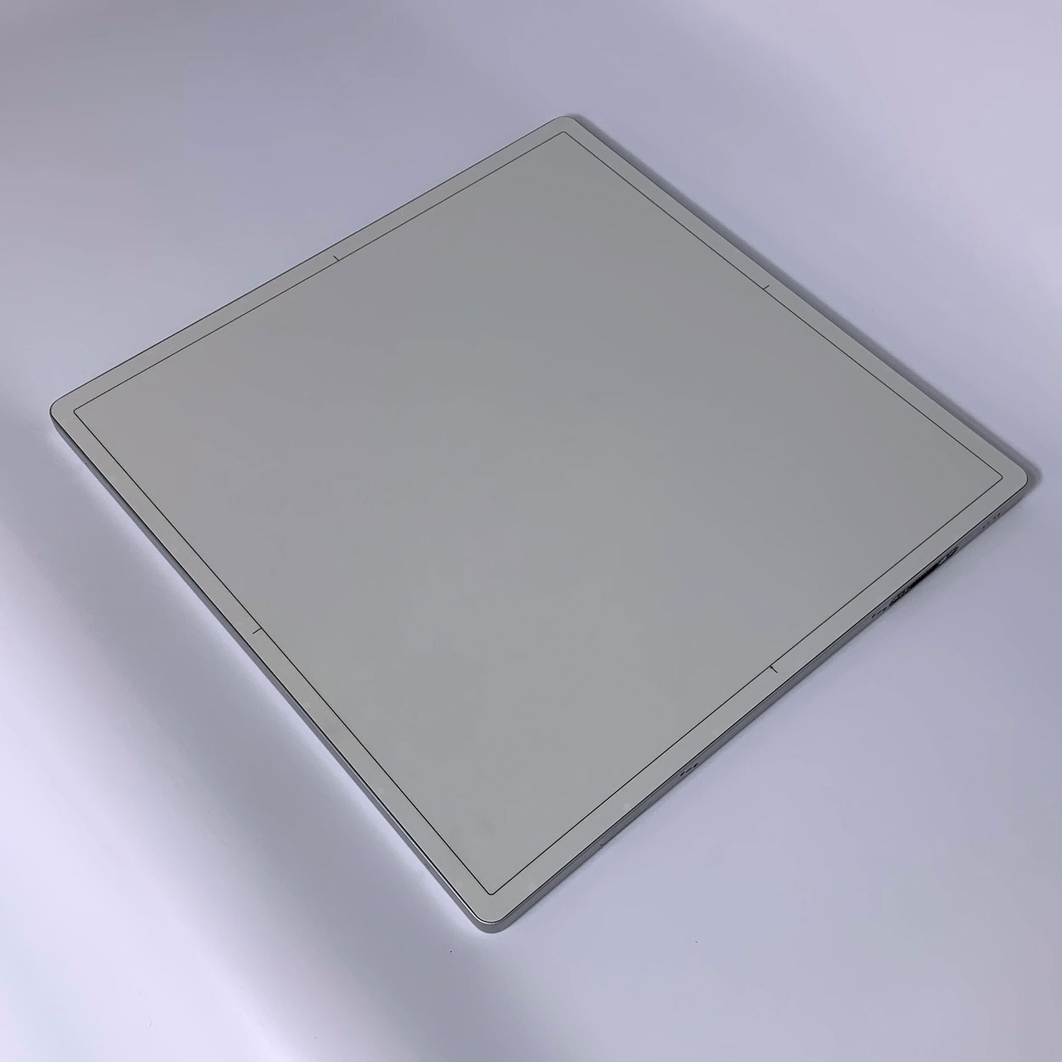
The World Health Organization estimated that 890 million adults were living with obesity in 2022, and radiography teams everywhere are feeling that shift in their daily caseload. What makes a bariatric examination different is not one single problem but several stacked ones — and most of them land on the image receptor:
One caution that applies throughout this article: BMI is a population-level surrogate, not a technique rule. It does not describe the thickness or tissue distribution of the specific body part being imaged. Positioning and exposure decisions belong to the attending radiographer and the facility’s validated technique charts — what this article covers is how to judge, before purchase, whether a detector is ready for this caseload.
Detector sizes are usually quoted by nominal cassette convention — 14×17, 17×17. But two panels with the same nominal label can expose different usable areas, because the active area inside the housing is what actually captures the image, and it is always somewhat smaller than the cassette outline. For bariatric work, where centimeters of coverage decide whether a chest or pelvis fits in one exposure, ask the supplier for the exact active area dimensions in millimeters — and compare, not just the brochure size.
A square 17×17-format panel covers a wide chest or pelvis in a single landscape-agnostic exposure, which is one reason square formats are often preferred in rooms that handle a broad mix of body habitus. A 14×17 panel works too, but orientation matters more: rotate it incorrectly and you clip anatomy. Our comparison of the two sizes covers this in detail; the bariatric-specific point is simpler — the more coverage per exposure, the fewer positioning holds you impose on a patient for whom holds are expensive.
For full-spine and long-leg studies, where even the largest single panel cannot cover the anatomy in one shot, the workflow becomes segmented: multiple exposures with planned overlap, stitched by software. Every stitch segment adds a breath-hold-and-hold-still interval — something larger patients often tolerate worst. If your caseload includes long-length studies on larger patients, ask the supplier specifically whether their acquisition software supports stitching, for which study types, and with what overlap requirements. Do not assume it is included; stitching capability varies by software package, not by panel size alone.
For a deeper dive on what a very large single active area (43 × 43 cm) changes about coverage, segmented studies and dose, see our dedicated large-format explainer — this article focuses on the patient side of that decision rather than repeating its numbers.
Cross-table and horizontal-beam laterals are among the most difficult bariatric views, because the detector must be held edge-on beside the patient — often with the patient’s tissue pressing against the panel housing. Ask how the panel is meant to be supported in that orientation: a dedicated holder, a wall-mount stand, or a table-side fixture. A panel that cannot be safely supported for horizontal-beam work quietly removes views from your protocol list.
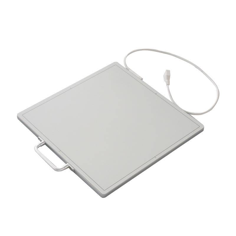
Supine abdominal and pelvic views put the panel directly under the patient — sometimes with a significant share of body weight bearing on the housing. This is where a set of mundane-sounding physical specs become clinical capabilities:
The cable-or-no-cable choice reads differently in bariatric work than in a general-room comparison. A wired panel’s cable must route around a patient who may occupy most of the table — a genuine trip-and-tangle hazard in tight quarters. A wireless panel removes the cable but adds a battery-management task. Neither is universally right; what matters is that the choice is made deliberately for this caseload. Our wired-versus-wireless guide walks the general trade-offs; for bariatric rooms, weight the routing question heavily.
Three detector-adjacent exposure topics matter more for larger patients, and all three belong in your pre-purchase questions:
A note on what we deliberately do not say: you will find no absolute dose numbers here, and no claim that any detector makes radiation harmless. Dose management is a facility protocol question governed by ALARA and local regulation; the detector’s job is to give your physicists and radiographers a stable, calibratable platform.
Before specifying a detector for a room that will regularly image larger patients, put these questions to the supplier in writing:
A supplier who answers these seven promptly, in writing, with numbers and documents, has almost certainly done bariatric-workable installations before. A supplier who answers with a catalog page has not — and your radiographers will discover that at the worst possible moment, with the patient already on the table.
Not necessarily. A square 17×17-format panel covers most chest and pelvis work in one exposure, and the large-format option mainly changes full-torso and segmented-study workflows. The right question is your caseload: which studies, on which patients, and where coverage currently forces retakes. Our large-format explainer gives a four-question checklist for that decision.
Yes, with attention to orientation. Landscape-versus-portrait placement matters more when anatomy is wider, and a clipped exposure is a repeat exposure. Confirm the exact active area first, then match the panel to the studies your room actually performs.
Technique factors generally rise with tissue thickness, but “much higher” is not a fixed rule and it is not primarily a detector specification. What the detector controls is repeatability: consistent calibration, standardized exposure index feedback, and image quality that does not tempt retakes. Dose decisions belong to your facility’s protocols under ALARA and local regulation.
The pairing of coverage and load: the exact active area in millimeters, and the panel’s rated load capacity with documentation. Those two numbers determine whether the routine examinations can be done in one exposure, safely, with the panel where it needs to be.
If you are specifying a detector for a room that serves a broad mix of body habitus, send us your study list and room layout. We will reply with the specific numbers — active areas, load ratings with documentation, handling specs — and where our standard configuration already covers them, in plain language, for your RFQ file.
Email: admin@newheek.cn | WhatsApp / Phone: +86 19062611512
Author:Newheek-Detector
Tel:+86 19062611512
Email:admin@newheek.cn
Company:Weifang Newheek Electronic Tech Co., Ltd.