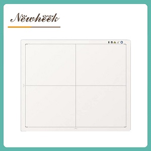
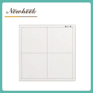
a large-format flat panel detector is a detector whose active imaging area is a 43 × 43 cm square — roughly 23% more area in every single exposure than the classic 35 × 43 cm rectangle. What it buys you is not sharpness (pixel pitch sets that) but coverage: chest and abdomen in one shot, fewer segmented spine and long-bone studies, fewer repositionings of the patient, and a lower cumulative dose per examination. A 2026 peer-reviewed study on large-format DR found examination times fell from 11.59 to 3.63 minutes and cumulative mAs dropped by about a third compared with conventional panels. Below we break down what a 43 × 43 cm field actually changes in daily work — and where it does not help.
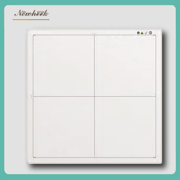
Flat panel detectors come in a small ladder of sizes, each inherited from a workflow: 10 × 12 inch panels for mobile and pediatric work, the 14 × 17 inch (35 × 43 cm) rectangle that has been the general-radiography standard since the film era, and the 17 × 17 inch (43 × 43 cm) square that has become the default on new fixed DR rooms. Above the square, a handful of manufacturers now offer extra-long formats around 43 × 120 cm, aimed at full-spine and full-leg studies.
One warning before comparing sizes: the inch figure is a nominal cassette size, and the active imaging area inside the housing is what matters. Within the square class alone, published active areas range from about 405 × 405 mm up to 430 × 430 mm — a difference you can see on any honest spec sheet.
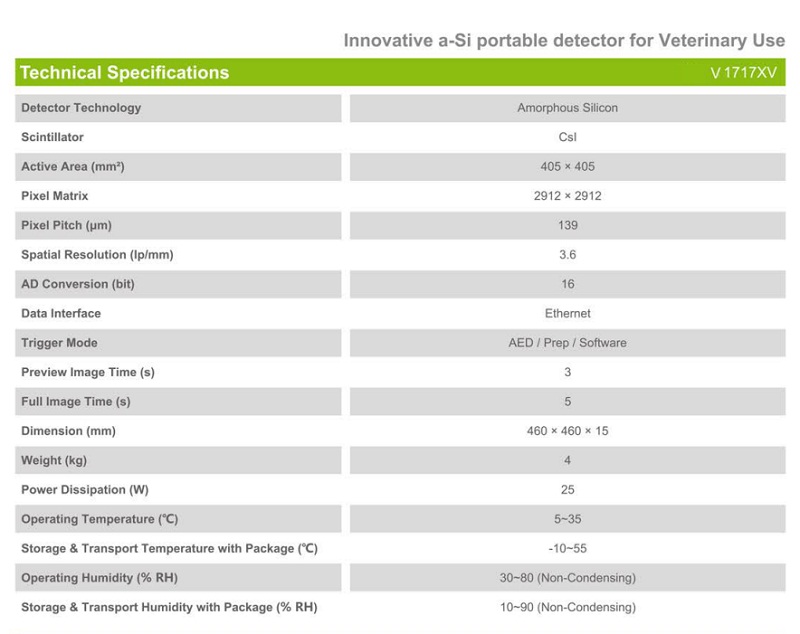
For reference, here is how our own published wired lineup compares across the two classes (full specifications in our detector specifications guide):
| Specification | 35 × 43 cm class | 43 × 43 cm class (large format) |
|---|---|---|
| Active imaging area | 350 × 430 mm | 430 × 430 mm |
| Area per exposure | 150,500 mm² | 184,900 mm² (+23%) |
| Pixel matrix | 2560 × 3072 | 3072 × 3072 |
| Pixel pitch | 139 μm | 139 μm |
| Spatial resolution | 3.6 lp/mm | 3.6 lp/mm |
| Acquisition time | 1 s | 1 s |
| A/D conversion | 16 bit | 16 bit |
| X-ray compatibility | 40–150 kV | 40–150 kV |
| Outer dimensions | 384 × 460 × 15 mm | 460 × 460 × 15 mm |
The square 43 × 43 cm field captures the chest and the abdomen of most adults without rotating the panel 90° between views. Compared with the 35 × 43 cm rectangle, each exposure covers about 23% more area (184,900 vs 150,500 mm²) — which converts directly into fewer exposures per patient and fewer opportunities for motion blur between shots.
Spine and long-bone work is where small panels hurt most: anatomy longer than the field must be taken in segments and stitched. A 2026 study cited in Frontiers in Medicine (Supakul et al.) reported that 16% of digital stitching examinations in a pediatric scoliosis cohort contained stitching errors — errors that carry real misdiagnosis risk. A larger field reduces how often you enter that workflow at all. (For the extremes of long-bone imaging, see the honest boundary section below.)
Every avoided repeat exposure is dose avoided. In the 2026 peer-reviewed comparison published in Frontiers in Medicine (Lin et al., 90 patients undergoing full-spine DR), the large-format workflow recorded cumulative mAs of 93.88 vs 146.89 for the conventional-panel workflow — roughly a third less radiation output per examination — alongside the time savings above. Industry analyses of long-format full-spine protocols (Browiner) report effective dose falling from 0.3–0.5 to 0.1–0.2 mSv for the same reason: fewer exposures, fewer retakes.
A common fear is that a bigger panel dilutes image quality. It does not: resolution is set by pixel pitch, the scintillator, and system MTF/DQE — not by field of view. Our square-format lineup keeps the same 139 μm pitch and 3.6 lp/mm resolution as the compact models, with the 3072 × 3072 matrix spanning the entire 430 mm width — so a femur imaged at the panel edge resolves just as well as one in the center. (For the layer under the pixels, see CsI vs GOS: Which Scintillator Should You Choose?)
Every eliminated panel rotation or repositioning is seconds saved per patient, repeated across a full list. In trauma, pediatric, and pain-limited patients the benefit is bigger than time: less manipulation of the injured area, fewer aborted attempts, and the 1-second acquisition window leaves less room for motion. This is the same mechanism behind the examination-time savings in the 2026 study — fewer steps, fewer repeats.
A 43 × 43 cm square is the most versatile single panel you can own. In general radiography it covers routine torso work; in veterinary imaging it handles large dogs and equine studies that force small panels into multi-shot sequences (the size is an established veterinary standard — see, for example, the Dormed VET-TECH DR4343, a 43 × 43 cm, 3072 × 3072 panel marketed specifically for animal imaging); in industrial NDT it inspects large castings and welds with fewer scans per part — which is precisely why market analysts (Mordor Intelligence) put large-size detectors at just over half of the flat panel market. For the veterinary angle in depth, see our veterinary detector buyer’s guide, and for industrial use, a-Si detectors for industrial NDT systems.
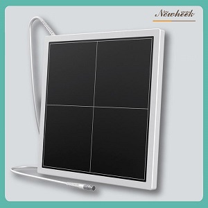
A 43 × 43 cm square is large-format, but it is not a long-format panel. Capturing an entire adult spine in a single exposure requires an extra-long detector around 43 × 120 cm — a format a few manufacturers have begun to offer, and which the 2026 Frontiers in Medicine study used (its large-format arm was a 42.58 × 120.87 cm panel). With a 43 × 43 cm square you will still segment a full spine into two or three exposures — just fewer of them, with less overlap guesswork, than a 35 × 43 cm rectangle requires. Judge any “single-exposure full spine” claim against the detector’s stated active area, and treat the long format as a separate purchasing decision.
Tell us your room type, your main anatomy mix, and whether the panel needs to be hand-carried — our product engineers will recommend a specific size and connectivity combination, wired or wireless (for that choice, see Wired vs Wireless Flat Panel Detector). Leave your details in the inquiry form on our Contact Us page and we will arrange a product manager to reply as soon as possible; you can also write to admin@newheek.cn or WhatsApp +86 19062611512 .
No. It covers about 23% more area per exposure, which pays off for torso, spine, and orthopedic workloads. For extremity-heavy or mobile workflows, the lighter and lower-cost 35 × 43 cm panel remains the practical choice.
No. Resolution is determined by pixel pitch, the scintillator, and system MTF/DQE. Our square-format and compact panels share the same 139 μm pitch and 3.6 lp/mm rating — compare those specifications between models instead of the size figure.
The square-format panel keeps the standard 460 × 460 mm full-size cassette footprint, so it fits installations designed for full-size cassettes. Verify your tray’s internal depth and clearance against the panel’s stated outer dimensions (about 460 × 460 × 15 mm) before ordering.
No. Single-exposure full-spine imaging requires an extra-long panel of around 43 × 120 cm. A 43 × 43 cm square reduces the number of segmented exposures compared with a 35 × 43 cm rectangle, but it does not eliminate them.
For a panel that lives in the table bucky or wall stand, wired GbE is simple and never needs charging. Hybrid models add 802.11ac wireless while keeping the wired path — useful if one panel must occasionally leave the room. The trade-offs are covered in Wired vs Wireless Flat Panel Detector.
No. The square format is an established veterinary standard for large-animal work, and in industrial NDT it reduces the number of scans per large casting or weld. The same panel serving a hospital radiography room can serve a clinic’s orthopedic list or a factory’s inspection bench.
Author:Newheek-Detector
Tel:+86 19062611512
Email:admin@newheek.cn
Company:Weifang Newheek Electronic Tech Co., Ltd.