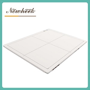
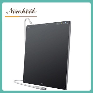
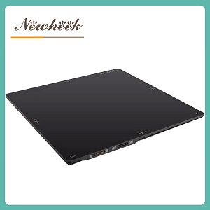
a flat panel detector is two layers stacked in one housing — a scintillator that converts X-rays into visible light, and a backplane that reads that light out as pixel data. Most “which detector is better” debates are actually happening in the backplane layer, where three technologies compete today: amorphous silicon (a-Si), IGZO, and CMOS. The short version: for static general radiography, a-Si remains the most proven and cost-efficient choice; for large-area high-speed dynamic imaging, IGZO is the rising option (20–30× the electron mobility of a-Si, 30–60 fps); for small-format, high-precision work like mammography and dental, CMOS leads on pixel pitch and readout noise. Below we explain what each backplane actually changes — with the numbers from 2026 market and conference data — and how to match one to your workload.
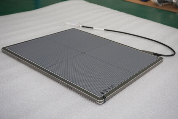
Start with the structure, because it prevents a common purchasing mistake. The scintillator (cesium iodide or gadolinium oxysulfide) decides how efficiently X-rays become light — we covered that choice in CsI vs GOS: Which Scintillator Should You Choose? The backplane — the thin-film transistor (TFT) or silicon layer directly under the scintillator — decides how completely and how quickly that light is converted into digital pixel values.
Because the two layers are independent, two detectors can both advertise “CsI” and still perform completely differently: same conversion layer, different readout layer. That is why any serious detector comparison must state the backplane technology — and why this article treats it as its own purchasing decision, not a footnote.
The figures below are drawn from the HDIN Research market report on medical X-ray flat panel detectors (2026–2031) and the SPIE Medical Imaging 2026 conference — not from any single vendor’s brochure:
| Property | a-Si (amorphous silicon) | IGZO (indium gallium zinc oxide) | CMOS (crystalline silicon) |
|---|---|---|---|
| Electron mobility | Under 1 cm²/V·s (HDIN) | Roughly 20–30× a-Si (HDIN) | Orders of magnitude higher — crystalline wafer (HDIN) |
| Practical pixel pitch | ~100–150 μm mainstream | Sub-100 μm (HDIN); migration toward 85–100 μm | Sub-50 μm (HDIN) |
| Full-resolution frame rate | Static workflows; low-speed dynamic | 30–60 fps (HDIN) | Up to 300 fps in specialized readouts (HDIN) |
| Large-area manufacturing | Mature; reliable yields at 43 × 43 cm (HDIN) | Same glass thin-film lines as a-Si (HDIN) | Wafer-limited; large areas need photolithographic stitching (HDIN, SPIE) |
| Relative cost | Lowest | List-price premium over a-Si (market analysts cite 25–35%), offset within roughly 24 months in total cost of ownership for fleet users | Highest at large sizes (SPIE: IGZO cheaper above ~17 × 17 inch formats) |
| Typical workload | Static general radiography; industrial high-dose NDT (Mordor Intelligence) | Large-area dynamic: fluoroscopy, dynamic DR, CBCT | Small-format precision: mammography, dental, surgical |
Amorphous silicon has carried clinical radiography for over two decades, and the reasons have not changed. It deposits on large glass substrates, which is why manufacturers can produce reliable yields at full 43 × 43 cm formats; it tolerates the high-dose duty cycles of industrial environments as well as the clinic; and it is the most cost-efficient backplane per square centimeter of coverage. Market analysts (Mordor Intelligence) still put a-Si at 36.8% of the digital radiography detector market in 2025 precisely because it remains the established operational standard for static work.
What is easy to miss is that a-Si keeps improving even without a new material name: market reporting points to pixel pitches migrating toward 85–100 μm, battery autonomy exceeding 15 hours on wireless panels, and better onboard automatic exposure detection. For a static radiography room — chest, abdomen, orthopedic lists — or a veterinary table, an a-Si panel remains the pragmatic default. Our current lineup across 14 × 17, 17 × 17, and veterinary portable formats is built on it, and the selection logic is covered in our detector specifications guide.
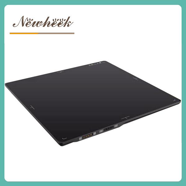
IGZO keeps everything that makes a-Si manufacturable — it deposits on the same large glass thin-film lines — but swaps the transistor channel material for a metal-oxide semiconductor with roughly 20–30× the electron mobility and extremely low off-state leakage (HDIN Research). The practical consequences are exactly what dynamic imaging needs: smaller transistors enable pixel pitches below 100 μm, lower noise improves signal-to-noise ratio, and full-resolution readout reaches 30–60 fps.
The durability question has also been answered. A peer-reviewed study covering 2024–early 2026 measured IGZO TFT arrays after 500 kGy of cumulative X-ray exposure and found spatial resolution maintained with dark current rising by less than 8% — a radiation-hardness result that outperforms older a-Si designs.
Most importantly for buyers, IGZO has crossed from novelty to product line. Varex launched 4343-format IGZO panels in 2025 claiming 40% lower image lag than equivalent a-Si models; Detection Technology extended its IGZO line across 1313, 3030, and 4343 formats in May 2026; and at SPIE Medical Imaging 2026, presenters argued IGZO holds a clear cost advantage over CMOS for panels larger than about 17 × 17 inches. Adoption is following: one major panel maker reported over 1,200 dynamic IGZO detectors shipped in a single half-year of 2025, and market trackers note IGZO dynamic panels rising to roughly 38% of new fluoroscopy and CBCT detector purchases in large Chinese cities — up from 12% two years earlier. Mordor Intelligence forecasts the CMOS/IGZO category to grow at 9.4% CAGR through 2031, the fastest of any detector technology.
A note on our own lineup: as of this writing, our published products are built on a-Si and CMOS backplanes. We describe IGZO here because it is reshaping what buyers should expect from the dynamic segment — not because we ship it. Treat any vendor’s IGZO claim, including future ones of ours, against the mobility, frame-rate, and dose figures above.
CMOS detectors are built on crystalline silicon wafers rather than glass, and that changes the physics: each pixel can carry its own amplifier, which minimizes readout noise and enables pixel pitches below 50 μm with frame rates up to 300 fps in specialized readouts (HDIN Research). That is why CMOS dominates high-precision, low-dose applications — full-field digital mammography, intraoral and dental CBCT imaging, and surgical imaging — where the active area is small enough to fit on a wafer.
The constraint is scale: tiling wafer segments into a large panel requires photolithographic stitching, which depresses yields and raises cost at large formats. This is why, per SPIE 2026 analysis, IGZO — not CMOS — has become the favored backplane for large-area dynamic systems. Within its natural format range, though, CMOS is unmatched: our own CMOS-based dynamic detector pairs a needle-like CsI scintillator with a CMOS sensor chip, delivering a 76 dB dynamic range, four gain levels for different application scenarios, real-time dynamic imaging at up to 60 fps at full resolution and full field of view, and calibration algorithms embedded in hardware — the kind of specification profile that only a wafer-based backplane supports.
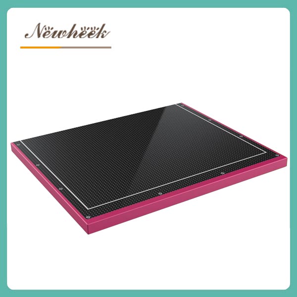
There is no universal “best backplane” — there is a best match for what your room does all day:
For how the backplane choice plays out in specific verticals, see our veterinary buyer’s guide (flat panel detectors for small veterinary clinics) and our industrial coverage (a-Si detectors for industrial NDT systems); for the connectivity half of the decision, start with Wired vs Wireless Flat Panel Detector.
Tell us your workload — static or dynamic, your anatomy mix, and your dose constraints — and our product engineers will recommend a detector combination that fits, rather than a catalog page. Leave your details in the inquiry form on our Contact Us page and we will arrange a product manager to reply as soon as possible; you can also write to admin@newheek.cn or WhatsApp +86 19062611512.
Not across the board. IGZO offers 20–30× the electron mobility, sub-100 μm pixel pitches, and 30–60 fps readout — decisive for large-area dynamic imaging. For static general radiography, a-Si remains the proven, lowest-cost standard, and its large-area yields at 43 × 43 cm are mature. Choose by workload, not by recency.
No. A static room’s workload — chest, abdomen, orthopedic exposures — does not benefit from high frame rates. An a-Si panel with a modern pixel pitch and good DQE, in the right scintillator, remains the cost-efficient answer; put the budget into the scintillator and size decisions instead.
CMOS achieves the smallest pixel pitches (sub-50 μm) and the lowest readout noise, which is why it dominates mammography and dental imaging. But at large formats it requires wafer stitching that raises cost and complicates yields — and sharpness in use is set by the whole chain (pixel pitch, scintillator, MTF/DQE), not the backplane name alone.
Yes, indirectly. Readout noise and DQE determine how much signal a detector can recover from a low-dose exposure; backplanes with lower noise and faster, cleaner readout hold image quality at lower dose. That is why pediatric and veterinary protocols weigh backplane-era specifications, not just size.
For most clinics, a-Si remains the practical choice: static exposures, robust duty cycles, and the best cost per area — with low-dose performance coming primarily from the scintillator and acquisition technique. High-frame-rate dynamic work is rare in veterinary practice; see our veterinary buyer’s guide for a full walk-through.
Yes — they are separate layers with separate supply chains. A CsI scintillator can be paired with a-Si, IGZO, or CMOS backplanes, which is why two “CsI” detectors can perform differently. Specify both layers explicitly in any purchase, and judge each on its own evidence.
Author:Newheek-Detector
Tel:+86 19062611512
Email:admin@newheek.cn
Company:Weifang Newheek Electronic Tech Co., Ltd.