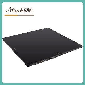
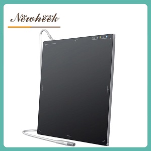
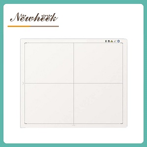
general radiography settles on two cassette sizes — 14×17 and 17×17 — because they cover chest, abdomen, spine and extremities in one plate. Mammography and industrial non-destructive testing (NDT) play a different game entirely. A mammography detector is a 24×30 cm cassette-scale panel resolved to 85 μm to catch microcalcifications; an NDT detector is a rugged 210×210 mm panel built to survive 320 kV and capture 60 frames per second. The sizes are not arbitrary — each one traces directly back to the physics of the task. Here is how to read them, using the three specialty detectors in our Other Size range as worked examples.
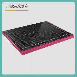
A mammography flat panel detector: 24×30 cm active area, 85 μm pixel pitch — dimensions inherited from the film-screen era, precision demanded by microcalcifications.
If you have read our comparison of 14×17 vs 17×17 flat panel detectors, you already know the logic: one rectangular plate must image everything from a hand to a full chest, film holders and wall buckies were built around those footprints, and so the DR market standardized on them. But step outside the general radiography room and that logic flips. In mammography, the anatomy is small, fixed and compressible — the job is not “cover the biggest area” but “resolve the smallest detail”. In NDT, the object is a weld, a casting or a pipe — the job is not anatomy at all, but defect visibility under extreme energy. The detector follows.
Three numbers define a mammography detector, and each has a reason.
Digital mammography kept the cassette dimensions of ISO 4090 film screens, so an FPD slides straight into the existing 24×30 cm bucky. That is also why a mammo panel is the classic retrofit part: the mechanical envelope of the machine stays, only the film gives way to electronics. Both detectors in our range — one amorphous-silicon based, one built on IGZO — use exactly this 24×30 cm active area for that reason.
Microcalcification clusters — an early marker in breast screening — can span well under a millimeter, so the detector must resolve structures far smaller than a general-radiography pixel (typically ~140 μm) would allow. At 85 μm pixel pitch, both panels reach 6 lp/mm spatial resolution, roughly double the fine-detail limit of a standard DR cassette. Resolution this fine is why mammography panels exist as a separate product line at all.
Breast tissue extends right up to the chest wall, and any detector edge that is thick or flat-tapered shadows that tissue out of the image. A tapered enclosure with a 1.5 mm (IGZO model) or 1.85 mm (a-Si model) slim edge pushes the active surface closer to the wall, extending field-of-view coverage where cancers hide. It is one of the least-advertised but most consequential specs on the plate.
Our range carries two mammography panels that share the 24×30 cm footprint and 85 μm pitch but diverge above it — a useful illustration that “same job” does not mean “same design”:
| Attribute | a-Si mammography sensor | IGZO mammography detector |
|---|---|---|
| Detector technology | Amorphous silicon | IGZO (oxide semiconductor) |
| Scintillator | CsI | CsI (direct deposition) |
| Active area | 240×300 mm | 240×300 mm |
| Pixel pitch | 85 μm | 85 μm |
| Pixel matrix | 2816×3528 | 3528×2816 |
| AD conversion | 14 bit | 16 bit |
| Data interface | GigE | 10G Ethernet |
| Intended use | FFDM, mag, biopsy — and tomosynthesis-capable | High-end FFDM and digital tomosynthesis, fast frame-rate read-out |
The pattern: a-Si paired with GigE is the proven, retrofit-friendly route (and handles tomography sequences); IGZO with a 10G interface and 16-bit conversion targets high-end FFDM and DBT, where faster read-out and wider dynamic range pay off. Why IGZO reads out faster is explained in our backplane comparison of a-Si vs IGZO vs CMOS; the short version is higher electron mobility enabling faster frame rates at low noise. And for a focused look at the breast-imaging workflow itself, see our piece on CsI and a-Si mammography detectors.
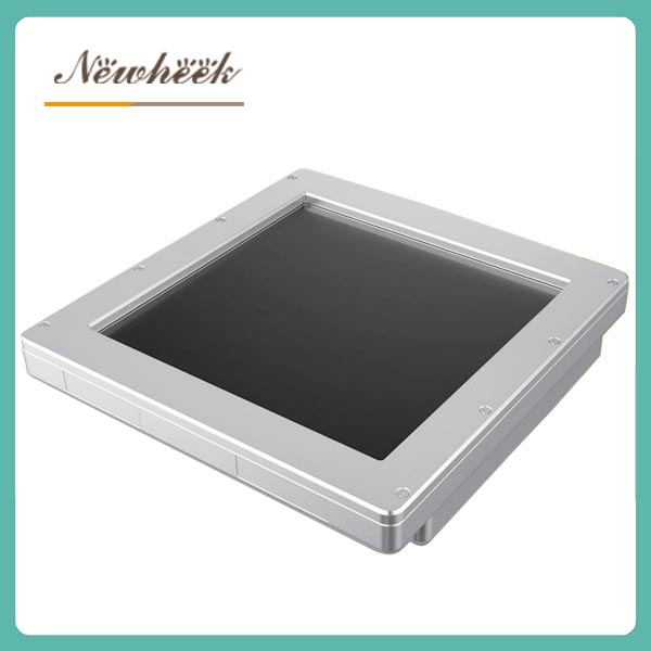
A flat panel detector for industrial NDT: amorphous silicon, 210×210 mm active area, rated for 320 kV high-energy radiation resistance.
The NDT panel ignores almost every mammography priority — and deliberately. Its job is watching welds, castings and assemblies under high-energy sources, often for hours of continuous inspection.
If you are weighing an NDT panel against a medical one for a mixed facility, the systemic differences are laid out in the NDT detector application category — see NDT X-ray detectors — alongside the medical line at digital mammography detectors and the full Other Size range.
| Attribute | Mammo (a-Si) | Mammo (IGZO) | Industrial NDT (a-Si) |
|---|---|---|---|
| Active area | 240×300 mm | 240×300 mm | 210×210 mm |
| Pixel pitch | 85 μm | 85 μm | 205 μm |
| Spatial resolution | 6 lp/mm | 6 lp/mm | 2.4 lp/mm |
| Scintillator | CsI | CsI, direct deposition | High-energy phosphor screen |
| AD conversion | 14 bit | 16 bit | 16 bit output |
| Interface | GigE | 10G Ethernet | GigE |
| Signature spec | 1.85 mm chest-wall edge, DBT-capable | 1.5 mm chest-wall edge, fast read-out | 320 kV tolerance, 60 FPS, <1% lag |
Read the table vertically and the design logic snaps into focus: the two mammo columns differ only in speed and depth (IGZO’s edge), while the NDT column differs in kind — bigger pixels, harder scintillator, exposure internals. Same underlying technology family, three completely different optimization targets.
Mammography inherited the ISO 4090 film-screen cassette format — 24×30 cm — so digital panels drop into existing buckies, and machines built for film can be converted to digital with a panel swap. The anatomy is smaller and fixed, so a larger plate would add nothing.
It enables 6 lp/mm spatial resolution — roughly double a standard DR cassette — which is the domain of microcalcification clusters, an early imaging marker in screening. Finer pitch is one of the defining differences between a mammography panel and a general radiography panel.
Breast tissue extends to the chest wall; a thick edge shadows it out of the field of view. Tapered enclosures with a 1.5–1.85 mm edge push the active area closer to the wall, improving coverage where lesions are most likely to appear.
Both panels in our range are tomosynthesis-capable, but they differ in headroom: the IGZO panel’s fast frame-rate read-out and 10G interface target high-end FFDM and DBT, while the a-Si panel with GigE covers FFDM, magnification and biopsy workflows. Confirm the frame-rate and acquisition sequence requirements of your specific DBT protocol before specifying.
NDT defect detection at typical source-to-object geometries is limited by source spot size and geometry before pixel pitch becomes the bottleneck. The panel’s priorities are high-energy radiation resistance (320 kV), frame rate (60 FPS), low image lag and integrator-friendly triggering and SDK access — which is exactly where its 205 μm / 210×210 mm design trades resolution for endurance and speed.
Specialty sizes reward buyers who start from the application: the anatomy, the defect, the energy, the sequence. If you are specifying a mammography retrofit, a DBT-capable panel or an NDT inspection line, contact us with the task and we will point you to the right panel and interface — including the full Other Size X-ray Detector range.
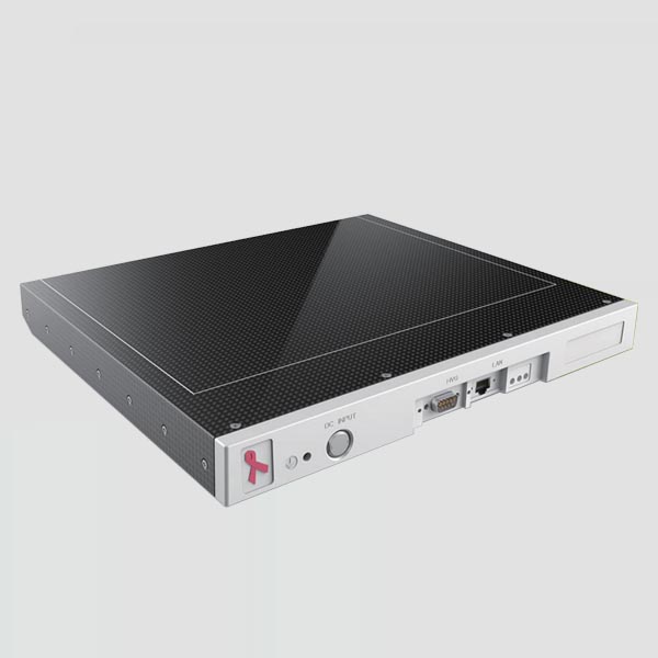
A mammography X-ray sensor with carbon-fiber front panel and slim chest-wall edge — the retrofit-friendly route into digital breast imaging.
Contact: admin@newheek.cn | WhatsApp +86 19062611512
Author:Newheek-Detector
Tel:+86 19062611512
Email:admin@newheek.cn
Company:Weifang Newheek Electronic Tech Co., Ltd.