Pediatric Radiography and the Detector: Sizes, Dose and Immobilization - Newheek DR Detector - Newheek DR Detector
Newheek specialize in the production of X-ray Detector.
Home›Blog ›Pediatric Radiography and the Detector: Sizes, Dose and Immobilization

Pediatric Radiography and the Detector: Sizes, Dose and Immobilization

children are not small adults, and most of what changes in pediatric radiography shows up at the detector. Three things differ from adult work: what “coverage” means when the patient is forty centimeters tall, how exposure is controlled — and how easily overexposure hides in a digital image — and how the panel itself is handled, held and cleaned around a small, moving patient. None of these require buying a special “pediatric detector,” but all three should change the questions you ask before you buy one. This article walks through each, with a pediatric add-on for your RFQ at the end.

Scope note: this is a buying-and-workflow guide, not a clinical protocol. Exposure factors, shielding practice and immobilization methods must be set by the radiologist and medical physicist for each room, in line with local regulations.

1. What Actually Changes When the Patient Is Small

An adult chest examination and a pediatric chest examination may use the same room, the same generator and even the same detector. What changes is everything around the exposure:

  • Coverage is easy; collimation is everything. A 14×17 panel more than covers an infant’s chest. The pediatric skill is imaging less of the child, not more — tight collimation to the clinical question is the single most effective dose-saving measure in published pediatric radiography guidance.
  • Exposure has less margin. Small bodies attenuate very little, so the exposure that produces a perfect image and the exposure that silently overexposes are close together — and digital processing makes both look fine on the screen.
  • The detector becomes handheld equipment. Around infants and toddlers, panels get held at odd angles, slid under squirming patients, grabbed by small hands, and disinfected between patients far more often than in adult routine.

Each of these maps to a concrete detector decision. Let’s take them in order.
2. Sizes: The Standard Cassettes Usually Win — Check the Details, Not the Label

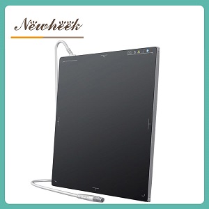
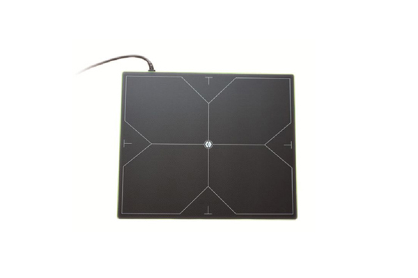
Compact custom-size flat panel detector with center cross-hair and wired connection

Here is the counterintuitive part: most pediatric radiography is done on the same 14×17 and 17×17 panels as adult work, because those sizes cover every pediatric body part with room to collimate. A dedicated small detector sounds appealing, but it splits your fleet, doubles calibration and QC work, and often sits unused as children grow into teenagers quickly.

That said, smaller formats do exist — compact 10×12-class panels and custom square footprints are common in specialty imaging (extremities, specialty suites), and some suppliers offer custom small sizes on request. If your pediatric workload is real and specific — a children’s hospital, a heavy orthopedic or NICU caseload — the questions to ask are about active area and pixel pitch, not the marketing size:

  • Active area, not cassette size. Two “14×17” panels can expose noticeably different active areas. For small patients, what matters is that the exposed area matches your collimated field — ask for the active area in millimeters and write it into the RFQ.
  • Pixel pitch trade-offs. Finer pitch resolves fine bony detail (valuable for non-accidental injury work and extremities), while coarser pitch generally buys dose efficiency. Published pediatric exposure charts treat this as a per-examination trade-off, not a universal rule.
  • Formats beyond the standard two are covered in more depth in our article on specialty detector sizes — including where 24×30 cm and compact industrial formats actually fit.

The practical buying rule: match the panel fleet to the department’s real case mix. A general hospital with occasional children does fine with standard cassettes and disciplined technique; a dedicated pediatric service should be pricing small-format options as a deliberate second unit, not a replacement.
3. Dose: The Digital Trap, and the Three Controls That Catch It

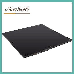
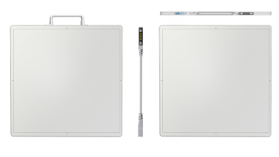
Multi-view technical drawing of a cassette-format wired detector showing front, edge thickness and connector side

Digital radiography gave pediatric imaging a gift and a trap in the same box. The gift: detectors respond to far lower exposures than film ever did, so dose can come down dramatically. The trap: post-processing makes an overexposed pediatric image look as good as a correctly exposed one. On film, overexposure was obvious. On DR, it is invisible — which is how “dose creep” happens one unnoticed exam at a time.

Published pediatric radiography guidance converges on three controls, all of which touch the detector:

  • Track a standardized exposure indicator. Use a detector whose exposure index follows a recognized standard (IEC 62494-1 defines the standardized EI), and review unusually high or low values as a routine QC habit — not to chase perfect numbers, but to catch drift. The indicator describes radiation reaching the detector, not patient dose, but it is the practical early-warning system your department has.
  • Treat automatic exposure control with respect in small children. AEC chambers were designed around adult anatomy. Pediatric positioning guidance — including ICRP publication 121 and the Image Gently materials summarized in published exposure charts — warns that in small children the anatomy may not reliably cover the chamber, causing premature or excessive termination. Many children’s hospitals use validated manual technique below a certain age and activate only chambers inside the collimated field. The buying consequence: the detector and system must support reliable manual technique and per-chamber AEC selection — confirm both, in writing.
  • Grid discipline. Grids improve contrast but demand more exposure. Published pediatric guidance (Image Gently and associated white papers) puts the crossover at roughly 10–12 cm of body thickness — below that, a nongrid technique is usually validated. Check how the detector performs in your nongrid pediatric protocols, not just in grid adult work.

One more point belongs here because parents ask about it constantly: shielding practice has shifted. Current consensus from medical physics organizations (AAPM and allied bodies, reflected in pediatric imaging alliances) recommends discontinuing routine patient contact shielding in diagnostic X-ray imaging — tight collimation protects better, and a misplaced shield can obscure anatomy (forcing a repeat) or interfere with exposure control. This concerns patient shields only; protective apparel for a caregiver who must hold a child remains a separate occupational-protection matter, and applicable law and facility policy always govern. If your team fields this question, the answer to give families is: the dose-control tools that matter are collimation, size-based technique and avoiding repeats.
4. Immobilization and Handling: The Detector as Handheld Equipment

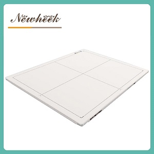
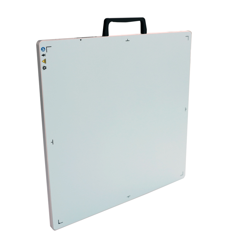
Cassette-format flat panel detector with carry handle, corner and center orientation markers

Motion is the enemy of pediatric radiography, and every motion-blurred image that forces a repeat is avoidable dose. Immobilization itself is a protocol question — positioning devices, caregiver holding with protective apparel, short exposure times — but it has a detector dimension that buyers overlook:

  • The panel gets held. For tangential views, swaddled infants, and wheelchair or seated positioning, a technologist or caregiver holds or braces the cassette. A molded carry handle, clear corner and center markers, and a weight a single gloved hand can control are pediatric-relevant features, not brochure decoration.
  • The panel gets grabbed. Anyone who has worked near toddlers knows objects get seized without warning. Drop and impact robustness is exactly the topic we covered in our durability article — for pediatric services, the “ask for the drop-test data and put it in the contract” advice stops being cautionary and becomes mandatory.
  • The panel gets cleaned, constantly. Between patients — and especially in NICU and isolation work — surfaces are disinfected far more often than in adult routine. Ask which disinfectant agents the housing and seals are rated for, in writing, and add it to the cleaning protocol.
  • Wired or wireless changes the choreography. A cable across a room where a child, a parent and a positioning device are all moving is a trip and yank hazard; a wireless panel removes the cable but adds battery and pairing discipline. We compared the two formats separately — for pediatric rooms, the honest answer is that the decision usually hinges on who holds the panel and where the patient is, not on the technology’s image quality.

5. The Pediatric Add-On for Your Detector RFQ

Whatever else your specification says, add these seven lines when children will be imaged on the unit:

  1. Active area in millimeters for each panel size offered — and the smallest formats available, if any.
  2. Exposure indicator standardized to IEC 62494-1, displayed after every exposure and exportable for QC review.
  3. AEC behavior at low exposure: per-chamber activation, chamber sensitivity settings, backup timer — and explicit confirmation that fully manual technique is supported with pediatric-appropriate minimum mAs.
  4. Weight, thickness and handle specification for every wireless and wired panel — the numbers a technologist’s wrist will live with.
  5. Mechanical robustness data: drop-test height, housing and seal ratings, stated per published test methods — or a written statement that the supplier does not test for it (which is also an answer).
  6. Approved cleaning and disinfectant agents for the housing, and the sealing method used at connectors.
  7. Pediatric processing presets or the ability to store them — size-based processing curves, if the acquisition software supports them. Do not assume presets exist; ask, and demo on a pediatric phantom if possible.

None of these seven lines requires a special pediatric product. They require a supplier who has thought about pediatric use — and the RFQ is where you find out who has.

Frequently Asked Questions

Do we need a separate small detector for pediatric patients?

Usually no. Standard cassette sizes cover pediatric body parts with room to collimate tightly, and collimation plus size-based technique does more for dose than panel size does. A small-format second panel earns its place in dedicated pediatric services with a specific case mix — price it as a deliberate addition, verify active area in millimeters, and check our article on specialty sizes for where compact formats genuinely fit.

Is a grid needed for children?

Follow your room’s validated protocol. Published pediatric guidance puts the typical crossover around 10–12 cm of body part thickness — below that, nongrid technique is usually validated because a grid demands extra exposure for little contrast benefit in thin bodies. The detector itself should hold up in your nongrid pediatric protocols; verify noise performance at low exposure rather than relying on grid work as the benchmark.

Should children still wear lead shields during an X-ray?

Current consensus from medical physics organizations recommends discontinuing routine patient contact shielding in diagnostic radiography: tight collimation is the more effective protection, and misplaced shields can hide anatomy (causing repeats) or interfere with exposure control. Always follow applicable regulation and facility policy, and explain to families that the real protections are collimation, size-based technique and avoiding repeat exposures. Protective apparel for a caregiver holding a child is a separate matter and remains standard practice.

Which detector spec matters most for pediatric dose?

Not a marketing number on the datasheet — a system behavior: a standardized exposure indicator you actually review, trustworthy manual technique with low minimum mAs, and honest AEC behavior at small patient sizes. Dose control in pediatric DR is a protocol-and-QC discipline built on those three detector behaviors, under ALARA principles and your medical physicist’s oversight.

Specifying a detector for a room that will image children? Ask us the seven questions above — we manufacture wired and wireless cassette-format flat panel detectors and will answer each line in writing. You can also reach us at admin@newheek.cn or WhatsApp +86 19062611512.

Related reading

Author:Newheek-Detector

(+86) 19062611512
admin@newheek.cn