the exposure index (EI) is a number that describes how much radiation actually reached the detector during one exposure. It is a measurement of the detector’s input — not a measurement of the dose the patient received, and not a pass/fail grade on anyone’s work. Used the right way, it is the cheapest dose-management tool a DR room owns: read as a trend, it exposes technique habits, flags a room that is quietly drifting toward routine overexposure, and gives you something concrete to fix before anyone gets hurt by it. Read as a single snapshot, it tells you almost nothing. This article explains the difference, retires three myths that cause most of the confusion, and lays out a one-hour monthly habit that keeps exposure discipline alive after the excitement of buying DR wears off.
Scope note: this article deliberately contains no absolute dose values and no vendor-specific EI formulas — manufacturers define and scale exposure indices differently, and quoting one vendor’s numbers against another’s is exactly the mistake this article is about. Ask your supplier in writing for their EI definition, target range and the reasoning behind it.

Here is a situation every imaging manager has walked into. Two radiographers share the same room. Same detector, same generator, same mix of patients. One of them gets exposure indexes that sit comfortably in the middle of the target range, month after month. The other’s numbers climb slowly, week after week, until somebody finally notices on a report.
Nothing is broken. No alarm is going off. The images from the second room often even look good — smooth, high contrast, easy to read. That is precisely why this drift goes unnoticed for so long: the room that is quietly overexposing produces the prettiest images in the building.
This is the first thing to understand about the exposure index: it is a mirror. It reflects what actually happened during the exposure — the technique that was set, the size of the patient, the habits of whoever pushed the button. When the number moves, it is usually telling you a habits story, not a hardware story. And a habits story is good news, because habits can be fixed for free.
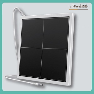
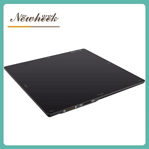
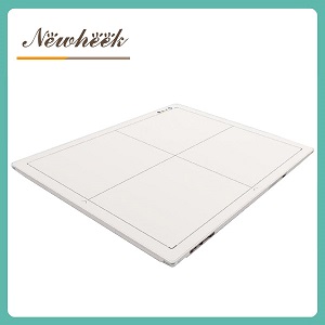
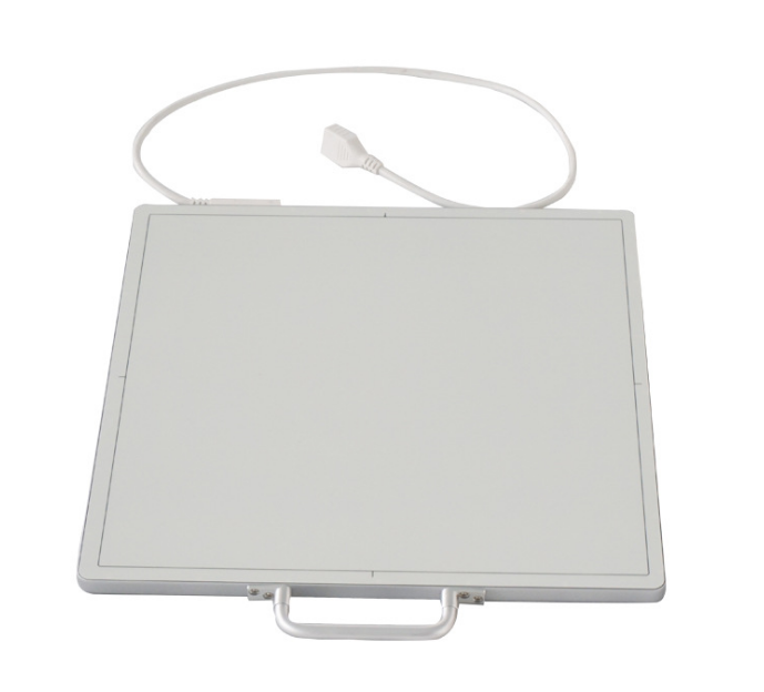
A flat panel detector is a remarkably honest device, but it is also a nearsighted one. It knows exactly one thing: how much radiation landed on it. It does not know how thick the patient was, which body part it was imaging, whether the beam was filtered and collimated perfectly, or whether the exposure was appropriate. It just measures what arrived.
Think of a rain gauge. It will tell you, precisely, how much rain fell into it. It cannot tell you whether you got wet on your way to the car. Patient dose is the “getting wet” part — it depends on beam quality, filtration, the body part, and things that happen outside the detector entirely. The exposure index is the rain gauge reading: a faithful record of the detector’s input and nothing more.
That is why the same exposure can produce a different EI on different equipment, and why no responsible engineer will convert an EI into “the dose this patient received” with a single formula. The number is a management tool, not a dosimeter.
A very low exposure index usually means the detector was starved — underexposed. The image comes out noisy and grainy, a radiologist questions it, and the exposure gets repeated. Now the patient has been exposed twice, and the repeat almost always uses a higher technique to make sure it “takes” this time. The lowest responsible dose is the one that produces a diagnostic image on the first exposure. Chasing the lowest possible EI is not dose discipline; it is a repeat-exposure factory.
Usually not. A gradually climbing EI most often means technique has crept upward — somebody nudged the settings, liked the result, and the room’s new normal was born. Genuine hardware problems (pixel defects, calibration drift, rising noise) announce themselves in QC and uniformity images long before they show up as an EI anomaly. If you suspect the panel and not the habit, our guide to artifacts, uniformity and calibration walks through what hardware drift actually looks like.
This is the one that causes cross-department arguments. Vendors scale and define their indices differently — some rooms display an EI, some a sensitivity-style value, some a deviation index that shows distance from target. The raw numbers are not comparable across manufacturers, across rooms, or sometimes even across protocols within one room. The only meaningful comparison is a number against its own detector’s target range, over its own history.
There is an uncomfortable truth from the CR-to-DR transition that every department should hear plainly: computed radiography was forgiving. Its wide exposure latitude meant that an overexposed plate still produced an acceptable image, so a room could run hot for years and nobody saw a reason to change. The error was invisible, so it persisted.
DR removes the invisibility. A digital detector records a huge range of signal faithfully, and the exposure index puts a number on every exposure. The technology now shows you exactly what your technique habits look like — but showing is not the same as lowering. If a room overexposed on CR, it will overexpose on DR, except now with a paper trail.
That paper trail matters more as imaging volumes grow. DR examination volumes worldwide keep climbing — China alone reports on the order of hundreds of millions of DR exams per year, according to Science Net’s 2026 coverage of UNSCEAR data — which is exactly why exposure discipline has moved from “nice to have” to a standard item in modern QC guidance. The visibility DR gives you is the tool. Discipline is what you do with it.
If you are still running a CR or film room and weighing the switch, our detector-side retrofit checklist covers what the panel side of that upgrade involves.
You do not need a physics department or a new software purchase to manage exposure well. You need a calendar slot and one honest hour a month. Here is the whole program:
That is the entire discipline. It costs one hour a month, it catches overexposure habits in weeks instead of years, and it produces exactly the documented, reviewed history that inspectors and accreditation visits increasingly ask to see.
Not all exposure-index implementations are equally useful, and the differences only surface if you ask before the purchase. Put these four questions in writing:
A vendor who answers these four with documents has built exposure management into the product. A vendor who answers with vague reassurance has not — and you will feel that difference every month, for the life of the room. When you are comparing detectors side by side, our spec-sheet guide explains how the other numbers on the datasheet connect to what you actually see in the exam room.
No. The EI measures the radiation that reached the detector during one exposure — the detector’s input. Patient dose depends on factors the detector never sees: beam quality, filtration, body part, collimation. The EI is a management and QC tool, not a patient dosimeter.
The target range defined by your detector’s manufacturer for your specific protocols — ask them for it in writing, including how it was derived. Raw values are not comparable between vendors or rooms; a number only means something against its own detector’s target and its own trend history.
Not the raw numbers. Different detectors scale their indices differently, and protocols and patient mixes differ too. Compare each room against its own history and its own target range — that is the comparison that produces honest, actionable answers.
Usually it means technique habits have drifted upward, which is a free fix. Genuine hardware problems — dead pixels, calibration drift, rising noise — show up in flat-field and uniformity QC images first. If you suspect the panel, run the QC checks before you touch the technique chart.
It can support lower-technique work, but it is not automatic and it is not free — image texture changes, and diagnostic confidence must be preserved. Treat it as a vendor question with an evidence requirement: documented comparisons at the reduced settings, on anatomy like yours, before anyone rewrites the technique charts.
If you are specifying detectors and want an EI definition, target range and export capability you can hold to, send us your room list and protocols. We will reply with the documentation behind our exposure reporting, in plain language you can file — and if a number in your monthly review ever needs a second opinion, we will help you read it.
Email: admin@newheek.cn | WhatsApp / Phone: +86 19062611512
Author:Newheek-Detector
Tel:+86 19062611512
Email:admin@newheek.cn
Company:Weifang Newheek Electronic Tech Co., Ltd.