DR detector system imaging process
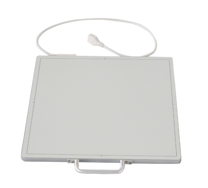
The ray transilluminated object reaches the DR detector (FPD), and the light is converted from the ray signal to visible light, focused by a thin film layer composed of photodiodes, and then sent directly to the computer for processing by the readout circuit to generate a digital image. The system includes: flat panel detector, transmitter/receiver (wireless or wired), X-ray machine, supporting computer and software.
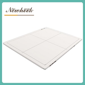
The DR detector is the core device of the DR system and contains a scintillator, which is directly coupled to the amorphous silicon (a-Si) thin film transistor (TFT) sensor, thereby generating high-pixel, highly sensitive digital images. It can produce images for a variety of objects.
The DR system has been applied in many non-destructive testing fields, such as welding, pipelines in cold storage, aerospace materials, high-voltage cable testing, etc.
The DR system also plays an unmatched role in the identification of archaeological cultural relics.
If you need a flat-panel DR detector or other radiology medical device, please contact us!
Author:肖恩