The flat panel detector is composed of a number of small squares, each of which is individually imaging, and the output digital signal represents the black and white value (grayscale value) of the small grid. This digital information is transmitted to a computer to form an X-ray image.
The gray value of each individual pixel in the X-ray image corresponds to each small square of the detector. For this reason, each detector cell is called a pixel.
It can be seen from this: the image quality of Dr Xray panel is determined by a single pixel.
Image pixel refers to the smallest unit composed of images composed of small squares, namely pixels. It exists as a single colored cell.
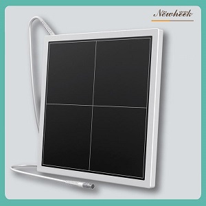
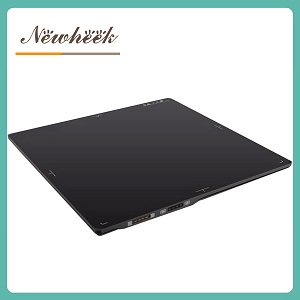
Newheek Dr Xray panel is divided into wired type and wireless type. Can meet your different purchasing needs.
Author:Glinda
Tel:+86 18953679166
Email:service@newheek.com
Company:Weifang Newheek Electronic Tech Co., Ltd.